
- H
- C
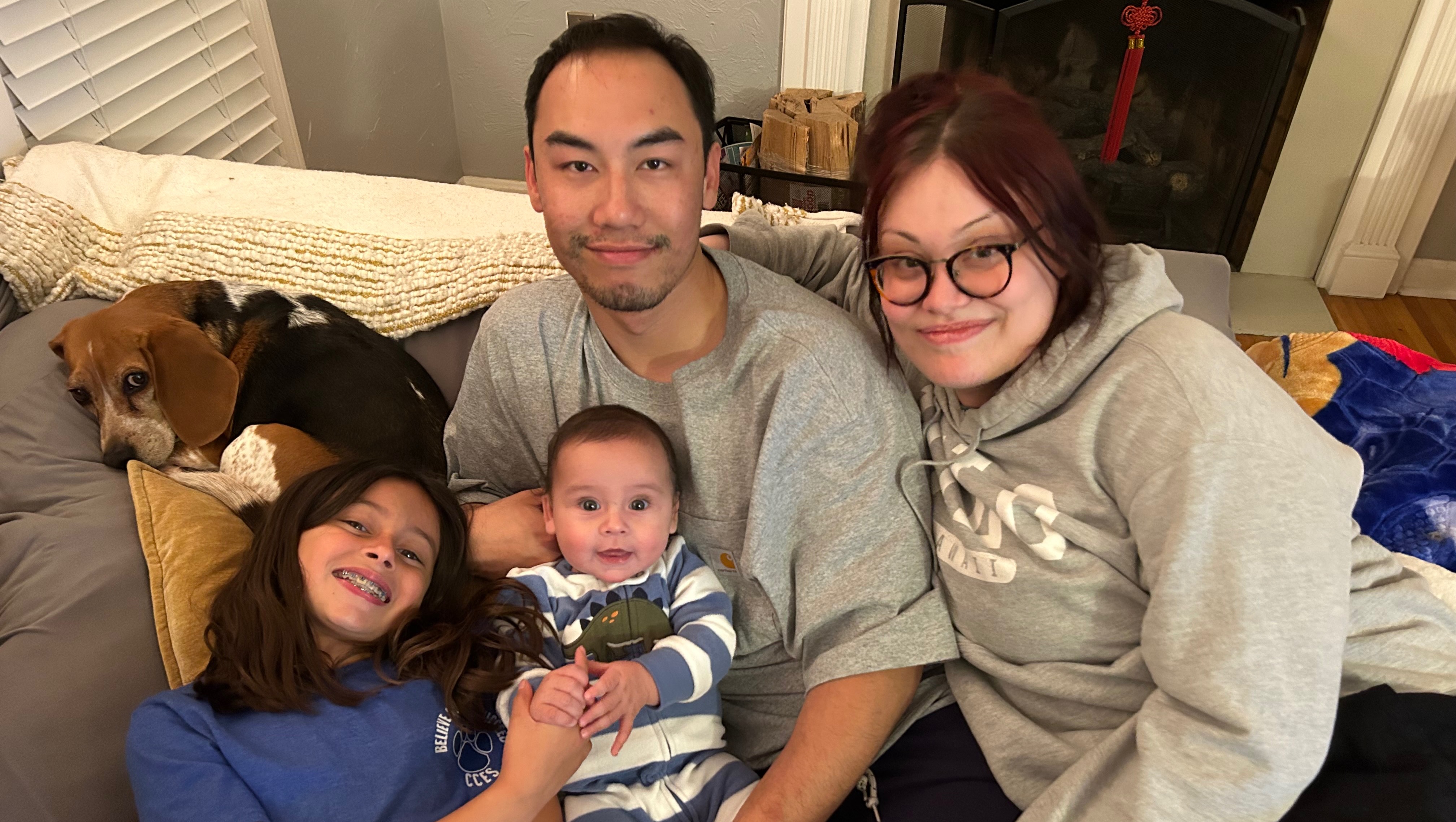
Hi everyone. For those of you who don’t know, my little sister Melissa Hale was diagnosed with stage 4 Osteosarcoma in March of this year at the age of 28. Her case is so rare that they have published it in a medical journal to study (see below). Not only does she have Osteosarcoma, but she also has Papillary Thyroid Cancer, and Anaplastic Thyroid Cancer. The cancer has spread to her brain, lungs, lymph nodes, as well as new masses growing in her throat area. She underwent a complete thyroidectomy, and has received radiation to the brain and neck region. Her next step will either be chemo or another form of cancer medicine which will hopefully be mostly covered by insurance.
In the past few weeks her health has taken a quick decline as she has not been able to swallow. She hasn’t been able to eat or drink, and is constantly coughing, gagging, nauseous, throwing up, and in overall pain. Fortunately she has been able to receive IV hydration to keep her fluid levels up, but she needs the calories to help stay strong. She will probably need a feeding tube soon to make that happen.
While all of this is happening in her body, she is unable to work and the debt keeps rising. Affording basic needs are becoming a struggle. So I am coming to you with open arms to please help in any way you can. Whether it’s $1 or $20. Your donations will help with:
- Rent and bills
- Transportation to and from hospital visits
- Basic household needs
- A mucus aspirator to help keep the phlegm out
- Holistic options
- Comfort care
- Medical expenses not covered by insurance
Her life partner, Gino has been carrying most of this weight financially, emotionally, and physically; and her family and friends have been an amazing emotional support system. I am here offering support with daily living tasks, but she still needs help financially. Melissa just wants to feel normal and healthy again. Thank you so much for any support you are able to offer, even if it’s just prayers and healing vibes.
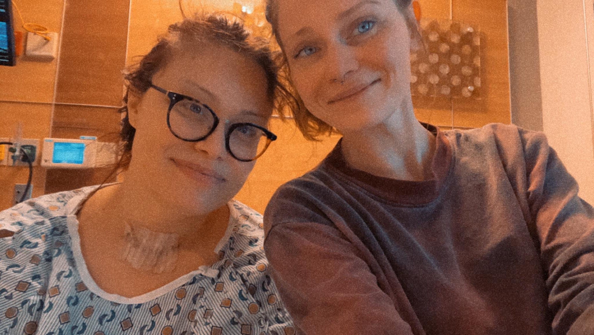
Update 7/5/23: I just wanted to update everyone on my sister’s progress. I didn’t want to jinx anything so i decided to wait until after today’s appointment. She had a CT scan done with contrast and met with the doctor about those results. Her neck area is definitely shrinking and her voice is almost back to normal. Her lung nodules are about the same but they haven’t grown so that’s great. She’s taking a cancer medicine that will help maintain/shrink all of the spots (brain, neck, and lungs). Her energy is starting to come back and slowly but surely she is finally able to eat. It was getting pretty scary for a while.
Thank you to everyone who has donated, called, text, messaged, mailed cards, and visited her. You have no idea how big of an impact you have all had. She has an amazing support system. It’s not over but she finally has some relief.


CASE REPORT:
A 28-year-old female presented to the clinic with a 1-year history of feeling a dull ache, pressure and progressive enlargement of her throat. She had no history of neck radiation, was euthyroid at the time, and had a maternal history of papillary thyroid cancer. She was instructed to get an ultrasound of her neck. Four nodules were detected, the most suspicious measuring 2.7 x 2.9 x 2.8 cm in the inferior pole of left thyroid lobe (Figs. 1 and 2). The nodule was nearly solid, hypoechoic, and lobulated or irregular borders, giving it a TI-RADS category 4. The patient followed up with a general surgeon and opted for total thyroidectomy.
Surgery began with an 8 cm cervical collar incision. As the sternohyoid and sternothyroid muscles were lifted, they were adherent to thyroid and surrounding soft tissues. There were multiple nodules of the left lobe that were thick and firm with mass effect, pushing the trachea to the right. Dissection of the thyroid was made using multiple techniques to separate the thyroid from the adherent soft tissue surrounding it, extending from the anterior to posterior aspects of the trachea and inferiorly past the clavicle into the substernal, upper mediastinal space.
The resected specimen revealed the left lobe of the thyroid that was almost completely replaced by anaplastic carcinoma, invading into the skeletal muscle, as well as widely throughout blood vessels and into the isthmus and vessels of the right lobe. The anaplastic tissue was surrounding a portion of the left lobe positive for papillary carcinoma (Fig. 3). Immunohistochemistry of the anaplastic carcinoma was positive for SATB2 and negative for ERG, CD31, CD34, desmin, MyoD1, myogenin, PAX8, TTF1 and thyroglobulin with Ki67 44.8 (Fig. 4). The papillary carcinoma was positive for PAX8, TTF1 and thyroglobulin, and negative for BRAFV600E and p53. There was involvement of 4 of 10 perithyroidal lymph nodes positive for anaplastic carcinoma without presence of papillary carcinoma.
Six days after the surgery, the patient collapsed in the bathroom after having visual changes and a feeling of intoxication. She was transported to the emergency department where bite marks were discovered on her tongue, but had no other visible signs of injury. CT of the head and chest was done which revealed hypoattenuating lesions in bilateral frontal lobes with mass-effect and without midline shift, a small pulmonary embolism, and multiple lung nodules. The patient was started on anticoagulation and instructed to follow up with oncology as an outpatient.
An F-18 FDG PET scan was completed which revealed multiple areas of increased uptake (Fig. 5). Cervical lymph nodes demonstrated hypermetabolism with maximum SUV of 9.8. There was multiple areas of hypermetabolism in the right lung, seen in the right upper, middle and lower lobes, with the greatest activity at the right hilum with an SUV of 22.3. There was no focal hypermetabolic lesion in the axial or appendicular skeleton. The patient underwent biopsy of suspicious lung nodules, which demonstrated metastatic papillary thyroid carcinoma in the right lower lobe, without osteosarcomatous components. Immunohistochemical stains demonstrated neoplastic cells positive for AE1/AE3 and PAX8, and negative for TTF1, thyroglobulin, and Napsin A and SATB2.
The oncology team made plans to start palliative radiation and chemotherapy. She was admitted for urgent initiation of radiation therapy after rapid tumor enlargement and CT showing possible mass invasion into the subglottic larynx and trachea. A core needle biopsy of the neck mass was taken showing tissue resembling previous osteosarcoma. She underwent daily radiation with 5 fractions of 300 cGy for 5 days in addition to oral temozolamide. The patient was discharged in stable condition without signs or symptoms of airway compromise.
Organizer and beneficiary
Melissa Lei'Lani Hale
Beneficiary