- A
- F
- L
Hello, everybody. My name is Jennifer Sandres and I am the mother to Sofia, who turned 14 in February. Sofia is in 8th grade at Carthage Jr. High and is in show choir, honor choir, she played volleyball, and is doing track and field. She is normally an A/B student, is very well-behaved, and has the best sarcastic and crude sense of humor a teen could have.
(En Español abajo)
In September of 2024, I took Sofia in to have her 6-month teeth cleaning, and while doing new x-rays, they noticed she had some teeth without roots. They told us to get her into an orthodontist and oral surgeon. We went to see Dr. LaFerla in Joplin. Dr. LaFerla said he had never seen a case like Sofia’s in his history of cases, except where there was very bad oral health. Sofia is cavity-free and has great oral health. He sent her to the oral surgeon after he and Dr. Wilson decided they believed there was a cyst in her gums. Dr. Bays did a CT scan and said there was a tumor in Sofia’s right maxillary sinus cavity. He said this was beyond his scope and we needed to go to Children’s Mercy Hospital in Kansas City, MO. We went to the ER in November of 2024 where they confirmed the mass and brought in oncology, ENT, oral, and facial surgeons from University Health. The next day, they did a biopsy orally at University Health since there were thoughts this was possibly a type of sarcoma cancer. The first biopsy came back inconclusive but showed there were giant cell clusters in a vascular background; possibly a Central Giant Cell Granuloma (CGCG). A second biopsy was performed and they still could not confirm anything but said it is compatible with CGCG. The doctors have decided to treat it as a CGCG.
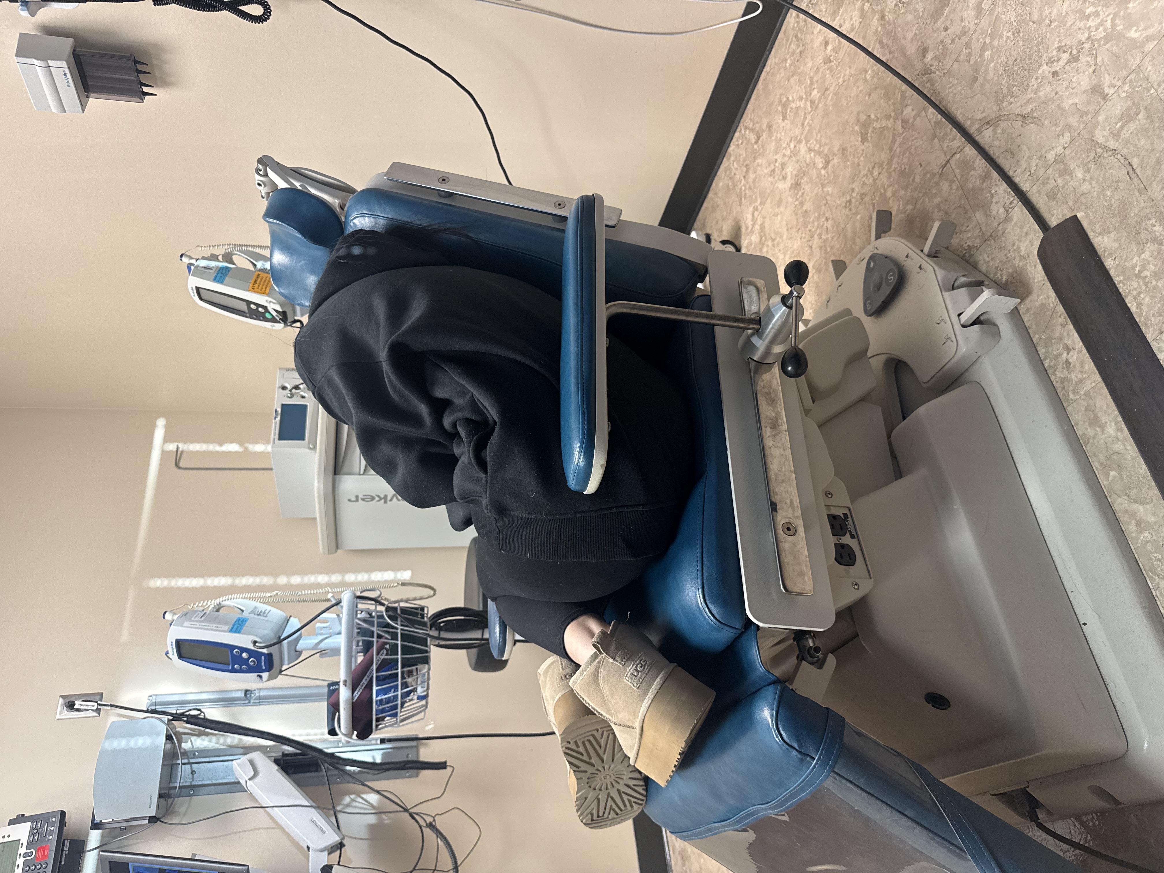
We have been making weekly trips to Kansas City, which is a 282-mile round trip for us. The plan is injections into the tumor every Tuesday with hopes that the tumor shrinks. Dr. Kim says there is a small chance the tumor would go away and then she would have to continue having scans until it came back as CGCGs are very recurring. Yesterday we had her 5th injection and they still can feel the mass. So, she needs one more injection and then two weeks after her last injection she will have a 3D x-ray so they can start planning her surgery. They said it’s the same as cancer and they will have to take 5 mm margins to help with the recurrence rate. We really don’t know much at this point, except she has a fairly large aggressive tumor, and surgery seems imminent. She will lose three teeth at minimum, with one already being gone; it was taken during the second biopsy. She also has a permanent hole into her sinus cavity from her mouth that has to be fixed from the second biopsy 3.5 months ago. She has bone damage and will need bone grafting. As I know more, I will keep this updated.
The money will be used to help with all the costs/bills we are incurring. She has health insurance and isn’t eligible for Medicaid. We have applied for travel help, etc., but because we have no confirmation on what the tumor is for sure, nobody will help with bills, grants, etc. Most people know I stay at home with Phoenix, who we adopted, and he is special needs. Luis works at Fletcher full time, but the car business has been super slow this year. We have been using our savings and have completely depleted them. We are now to the point of going without paying bills to get Sofia to and from KC, her meds, keeping her bills paid, etc. The funds will be used to pay for our gas and food to KC. Buy Sofia meds. Keep her hospital and Dr bills paid up as much as possible. She had a $3000 deductible on one health insurance and $750 on the other. She has $1000 yearly pay out and $1500 on her other dental plan. She will need implants as well.
We also tried getting her in at Mayo Clinic but never heard back from them after sending all her medical records to them.
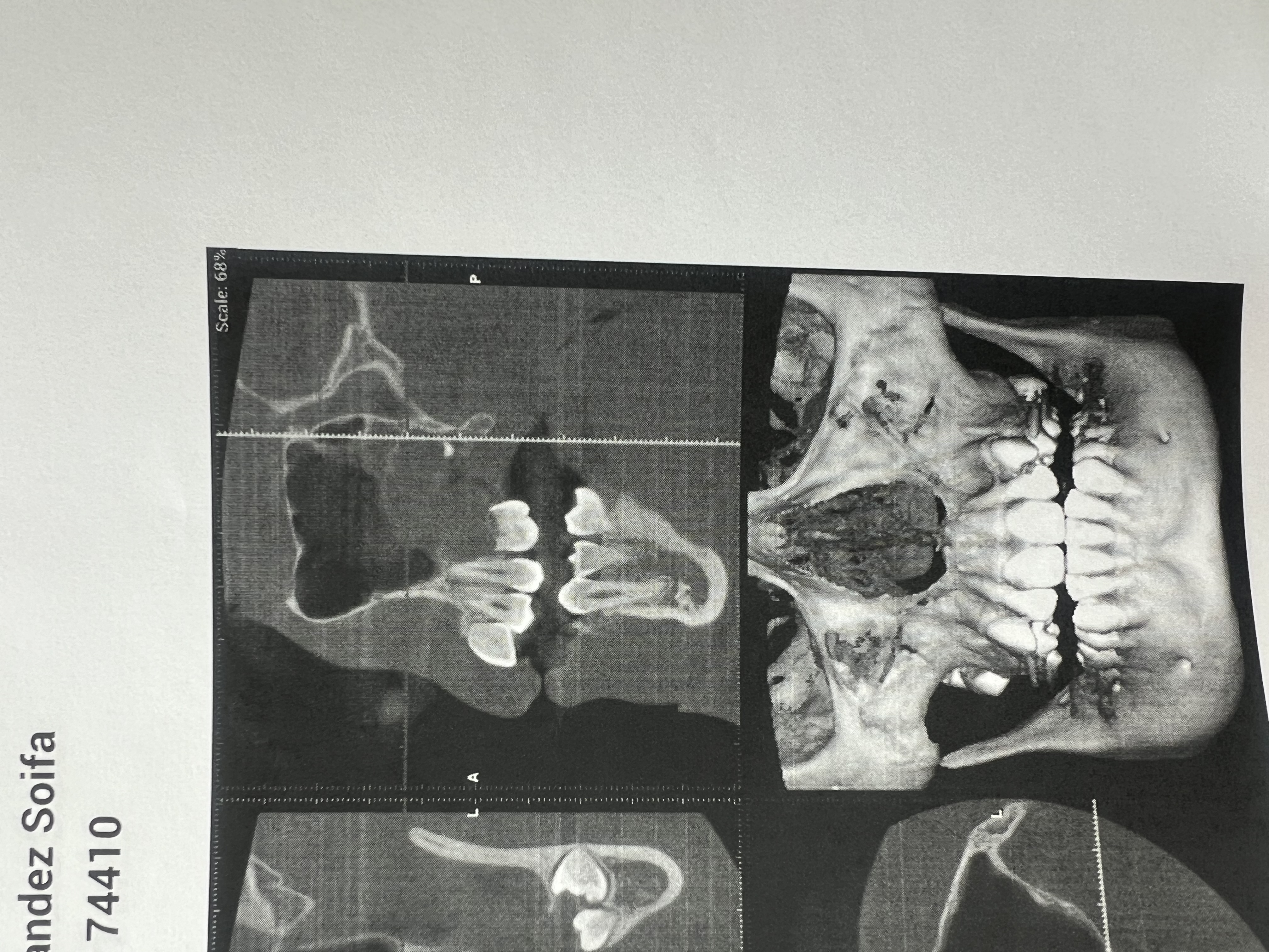
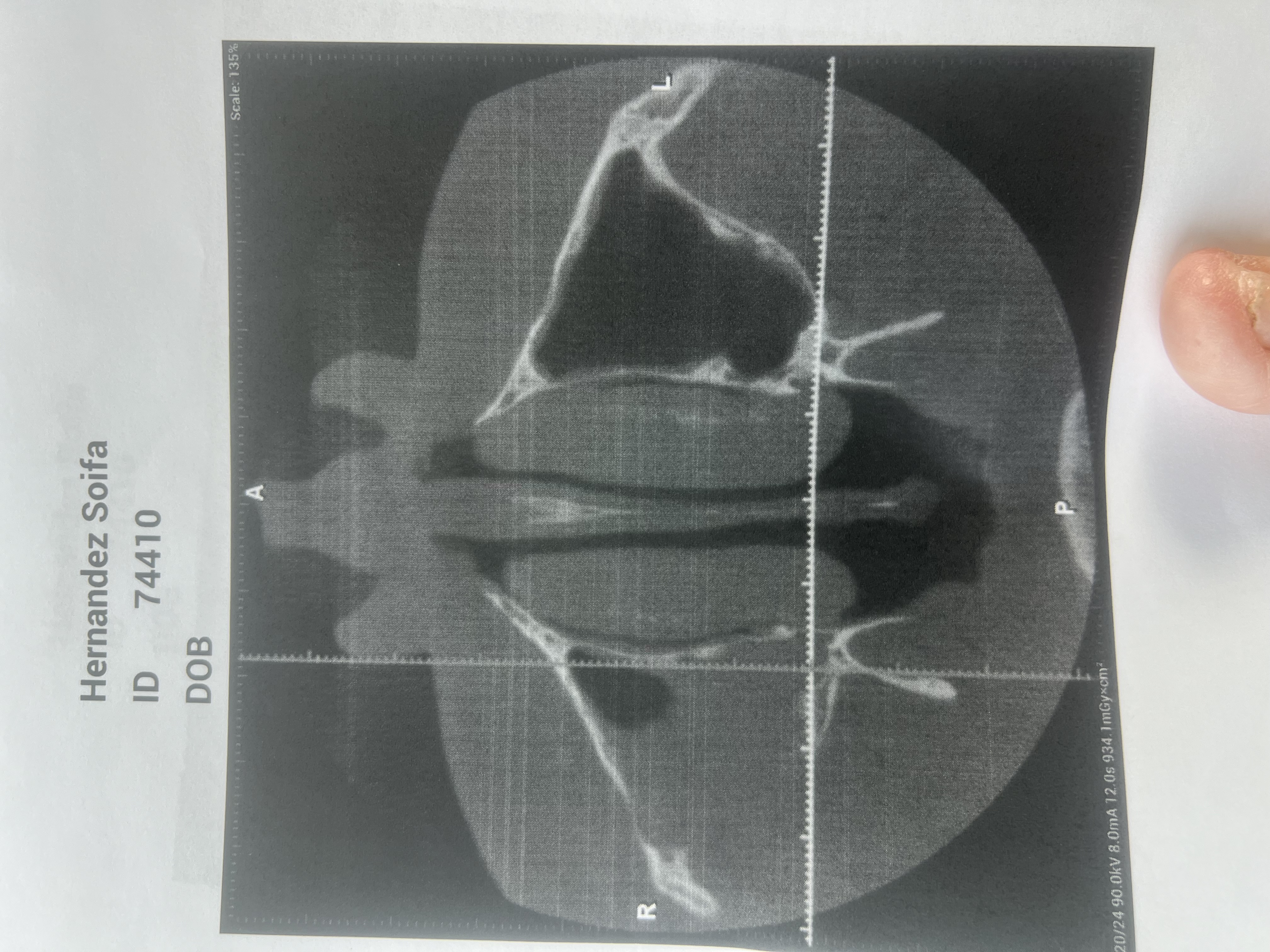
CT Findings with Dr Bays “X-ray: CBCT shows 2.5-3 cm expansile mass in the right maxillary sinus involving teeth #1, 2, 3. There has been root resorption of #1, 2, 3. There is erosion of the lateral and posterior wall of the maxillary sinus as well as the hard palate and lateral nasal wall in the #2-3 area. It extends near to the orbital floor laterally.”
Children’s Mercy CT Findings “There is an approximately 2.2 cm AP x 2 cm TR x 2.8 cm oblique SI soft tissue mass centered in the inferior right maxillary sinus. There is erosion of the surrounding maxillary walls laterally, posteriorly, inferiorly and inferomedially, invading the lamina dura of the first, second, and third molars. There is associated smooth erosion of the roots of these molars, with some preservation of the anterior of the first molar. There is associated severe thinning of the nasal surface of the maxilla, but without definite complete breech into the inferior nasal meatus. This superior margin of the mass approaches the lateral orbital floor and inferior ophthalmic vein canal, but does not appear to breech them.
The orbits and globes are normal. No maxillofacial fracture is present. There is a small 0.6 cm sclerotic focus in the right anterior mandible, possibly a bone island. Otherwise, the mandible and temporomandibular joints are normal.
The mastoid air cells are clear. There is a small mucous retention cyst in the superior left maxillary sinus, and there is mild scattered caudal thickening in the remaining paranasal sinuses.
The imaged intracranial anatomy is normal.
IMPRESSION:
1. Erosive soft tissue mass centered in the right maxilla, measuring up to 2.8 cm.
2. Associated destruction of the maxillary sinus walls and roots of the first-third molars.”
Hola a todos. Mi nombre es Jennifer Sandres y soy la mamá de Sofía. Sofía es la hija de Oswaldo Velazco y hijastra de Sandra (Pérez) Velazco y Luis Sandres ella es un estudiante de Carthage y tiene 14 años. En Septiembre del año pasado llevamos a Sofía con la dentista para un limpieza. Cuando ellos saco las radiografías se dieron cuenta que había tres dientes sin raíces. El dentista mandó a ella con otro doctor en Joplin. Ese doctor mandó a Sofía a un doctor medical q puede operar de la cara y en la boca. Ese doctor se dio cuenta q había un tumor agresivo en el seno nasal del maxilla de Sofía. Ese doctor nos mandó para Kansas City y de ahí ellos nos mandó a otro hospital. Ella ha tenido dos biopsias pero no pueden confirmar exactamente q es el tumor. Ello piensan q es CGCG (Central Giant Cell Granuloma) agresivo. Ahorita ella tiene 5 semanas recibiendo tratamientos semanales y falta una semana más. Entonces tenemos q viaje a Kansas City cada semana y es un viaje de 282 millas de ira y venida. Entonces tenemos tres semanas más van a sacar otro resonancia para ver el tumor y hacer los planes para remover el tumor. Va a ser un operación extensivo donde tiene q sacar 5 mm de los huesos etc. Estamos juntado el dinero para los viajes y biles q va a quedar después de seguro medical.
"Se observa una masa expansiva de 2,5-3 cm en el seno maxilar derecho que afecta los dientes 1, 2 y 3. Se ha observado reabsorción radicular en los dientes 1, 2 y 3. Se observa erosión de las paredes lateral y posterior del seno maxilar, así como del paladar duro y la pared nasal lateral en la zona 2-3. Se extiende lateralmente cerca del suelo
orbitario."
"Se observa una masa de tejido blando en la porción distal del maxilar inferior derecha de aproximadamente 2,2 cm AP
x 2 cm TR x 2,8 cm oblicua. Se observa erosión de las paredes maxilares circundantes lateral, posterior, inferior e inferomedialmente, que invade la lámina dura del primer, segundo y tercer molar. Se observa erosión suave asociada de las raíces de estos molares, con cierta preservación de la parte anterior del primer molar. Se observa un adelgazamiento severo de la superficie nasal del maxilar, pero sin una ruptura completa definida hacia el meato nasal inferior. Este margen superior de la masa se aproxima al suelo orbitario lateral y al conducto venoso oftálmico inferior, pero no parece romperlos. Las órbitas y los globos oculares son normales. No se observa fractura maxilofacial.
Se observa un pequeño foco esclerótico de 0,6 cm en la mandíbula anterior derecha, posiblemente una isla ósea. Por lo demás, la mandíbula y las articulaciones temporomandibulares están Normal. Las celdillas mastoideas están limpias. Se observa un pequeño quiste de retención mucosa en el seno maxilar superior izquierdo y un leve engrosamiento caudal disperso en los senos paranasales restantes. La anatomía intracraneal en la imagen es normal. IMPRESIÓN: 1. Masa erosiva de tejido blando centrada en el maxilar derecho, que mide hasta 2,8 cm. 2. Destrucción asociada de las paredes del seno maxilar y las raíces de los primeros y terceros molares."