

Help Maigret Peña win the battle against breast cancer
0% complete
$535 raised of
11 donations
We are raising funds for my mother's cancer treatment
We need help with her oncologist appointment to understand what will be the next steps for her treatment.
We offer no raffles, sweepstakes, giveaways or promotions for this GoFundMe.
This is what we've done so far!
✅ - First Biopsy
✅ - Mammography
✅ - Mammary Eco
✅- Thorax Xray
✅- Abdominal Eco
✅- Pelvic Eco
✅- Blood Work
✅- Oncologist appointment
What we havent so far!
❌ - 3 CT scans 250
❌ - 1 PET Scan 250
Appointment results
We saw the oncologist surgeon who gave us an updated budget and treatment plan. We will be working with the medical oncologist for now until the tumor has decreased in size enough for the surgery. So we need roughly 8 cycles of chemotherapy which would be 400 each, but we are being advised that through some public means we can get it for free, that way we'd only have to save up for the surgery and radiotherapy.
We will update this plan again after the next appointment.
Break down cost of next appointments:
Oncologist Appointments: 77$ each
We do not know how often we will need to check in but we know how much it will be for each appointment
Tumor Removal: 3,300$usd
If the tumor responds to the chemo how we want it to.
Second Biopsy for Tumor Removal: Roughly 400$
The tumor is currently 2 cm and we are working against the clock to raise enough funds to save her life. After the next appointment we will keep updating.
We are currently residing in Venezuela and the funds will be transferred to my boyfriend’s bank account who lives in the US. He will transfer 100% of the funds to us through Zelle and PayPal as we require them for her appointments.
Documents:
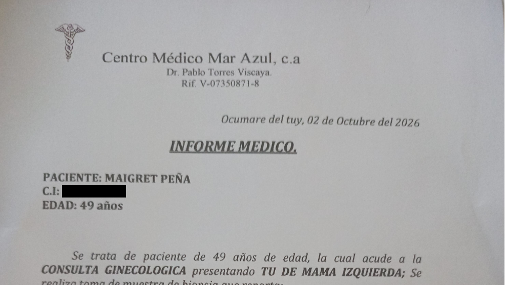
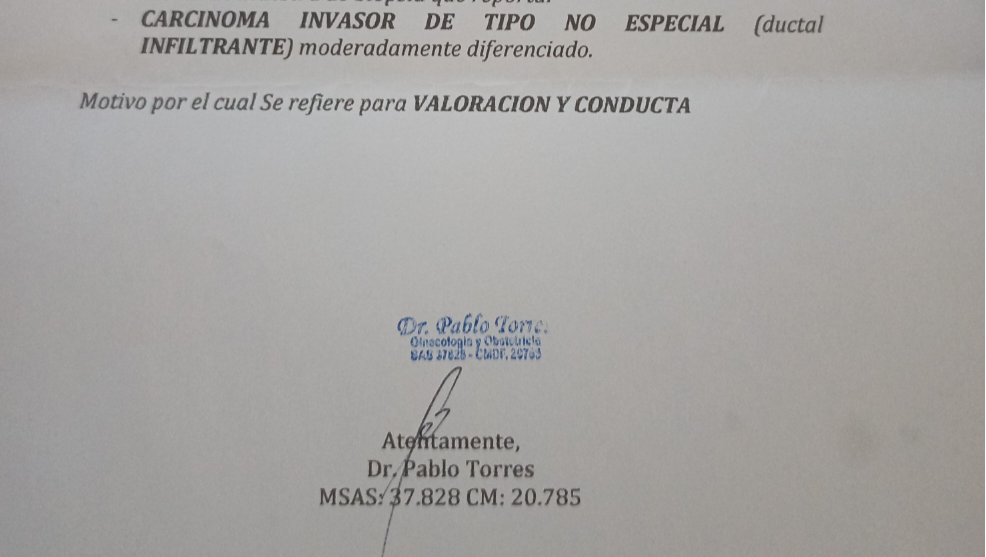
Spanish Transcript
Paciente: Maigret Peña
C.i: [CENCURADO]
Edad: 49 años
Se trata de paciente de 49 años de edad, la cual acude a la CONSULTA GINECOLÓGICA presentado TU DE MAMA IZQUIERDA; Se realiza toma de muestra de biopsia que reporta:
CARCINOMA INVASOR DE TIPO NO ESPECIAL (ductal INFILTRANTE) moderadamente diferenciado.
Motivo Por el cual Se refiere para VALORACIÓN Y CONDUCTA
English Transcript
Patient: Maigret Peña
ID No.: [REDACTED]
Age: 49 years
The patient is a 49-year-old female presenting for a gynecological consultation with a left breast tumor. A biopsy was performed, yielding the following report: Moderately differentiated invasive carcinoma of no special type (infiltrating ductal carcinoma). Reason for referral: Evaluation and management.
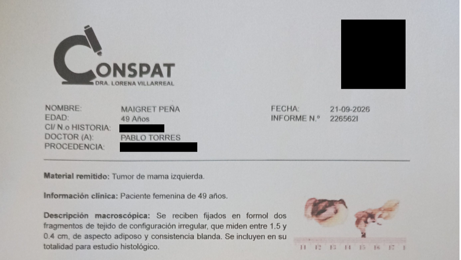
Spanish Transcript
Nombre: Maigret Peña
Edad: 49 Años
CI/ N.o História: [CENSURADO]
Doctor (A): Pablo Torres
Proceddencia: [CENSURADO]
Fecha 21-09- 2026
Informe N: 2265621
Material remitido: Tumor de mama izquierda
Información clínica: Paciente femenina de 49 años
Descripción macroscópica: Se reciben Fijados en formol dos fragmentos de tejido configuración irregular, que miden entre 1.5 y 0.4 cm, de aspecto adiposo y consistencia blanda. Se incluyen en su totalidad para estudio histológico
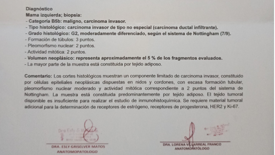
Diagnostico
Mama izquierda; biopsia:
-Categoria B5b: Maligno, carcinoma invasor.
-Tip histógico: carcinoma invasor de tip no especial (carcinoma ductal infiltrante).
Grado histológico: G2, moderadamente diferenciado, según el sistema de Nottingham (7/9).
-Formación de túbulos: 3 puntos.
-Pleomorfismo nuclear: 2 puntos.
-Actividad Mitótica: 2 puntos.
-Volumen neoplásico: representa aproximadamente el 5 % de los fragmentos evaluados.
-La mayor parte de la muestra está constituida por tejido adiposo.
Comentario: Los cortes histológicos muestran un componente limitado de carcinoma invasor, constituido por células epiteliales neoplásicas dispuestas en nidos y cordones, con escasa formación tubular, pleomorfismo nuclear moderado y actividad mitótica correspondiente a 2 puntos del sistema de Nottingham. La muestra está constituida predominantemente por tejido adiposo. El tejido tumoral disponible es insuficiente para realizar el estudio de inmunohistoquímica. Se requiere material tumoral adicional para la determinación de receptores de estrógeno, receptores de progesterona, HER2 y Ki-67.
Dra Esly Griselver Matos
Anatomopatologo
Dra Lorena Ve Larreal Franco Anomatólogo
English Transcript:
Name: Maigret Peña
Age: 49 years
ID/Medical Record No.: [REDACTED]
Doctor: Pablo Torres
Origin: [REDACTED]
Date: 21-09-2026
Report No.: 2265621
Specimen submitted: Left breast tumor
Clinical information: 49-year-old female patient
Macroscopic description: Two formalin-fixed tissue fragments of irregular configuration, measuring between 1.5 and 0.4 cm, are received; they have an adipose appearance and soft consistency. They are entirely submitted for histological study.
Diagnosis
Left breast; biopsy:
-Category B5b: Malignant, invasive carcinoma.
-Histologic type: Invasive carcinoma of no special type (infiltrating ductal carcinoma).
Histologic grade: G2, moderately differentiated, according to the Nottingham system (7/9).
-Tubule formation: 3 points.
-Nuclear pleomorphism: 2 points.
-Mitotic activity: 2 points.
-Neoplastic volume: represents approximately 5% of the evaluated fragments.
-The majority of the sample consists of adipose tissue.
Comment: Histological sections show a limited component of invasive carcinoma, consisting of neoplastic epithelial cells arranged in nests and cords, with scant tubule formation, moderate nuclear pleomorphism, and mitotic activity corresponding to 2 points on the Nottingham system. The sample consists predominantly of adipose tissue. The available tumor tissue is insufficient to perform immunohistochemical analysis. Additional tumor material is required for the determination of estrogen receptors, progesterone receptors, HER2, and Ki-67. Dr. Esly Griselver Matos
Pathologist
Dr. Lorena Ve Larreal Franco
Pathologist
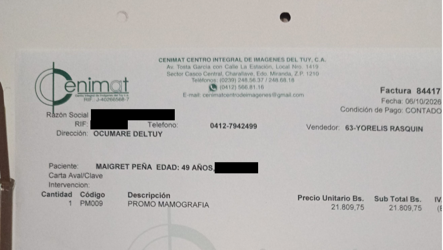
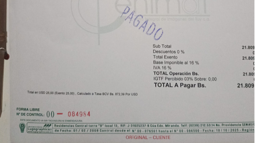
Spanish Transcript:
Factura 84417
Fecha 06/10/2026
Condición de Pago: Contado
Razón Social: [CENSURADO]
RIF; [CENSURADO]
Telefono: 0412-7942489
Vendedor: 63-Yorelis Rasquin
Dirección: [CENSURADO]
Paciente: Maigret Peña Edad; 49 Anos, 12614255
Carta Aval/Clave
Intervencion
Cantidad: 1
Codigo: PM009
Description: ROMO MAMOGRAFIA
Precio Unitario Bs. 21,809.75
Sub Total Bs: 21,809.75
English Transcript
Invoice 84417
Date: 06/10/2026
Payment Terms: Cash
Company Name: [REDACTED]
Tax ID (RIF): [REDACTED]
Telephone: 0412-7942489
Salesperson: 63-Yorelis Rasquin
Address: [REDACTED]
Patient: Maigret Peña
Age: 49 Years, 12614255
Letter of Guarantee/Code
Procedure
Quantity: 1
Code: PM009
Description: MAMMOGRAPHY PROMO
Unit Price (Bs.): 21,809.75
Subtotal (Bs.): 21,809.75
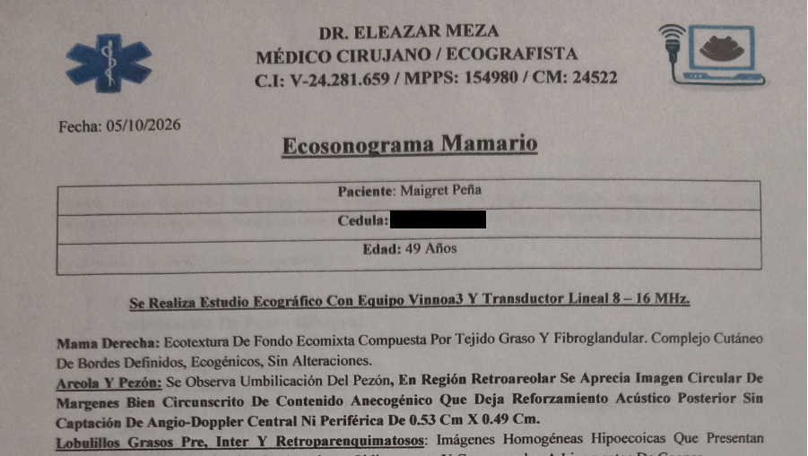
Spanish Transcript:
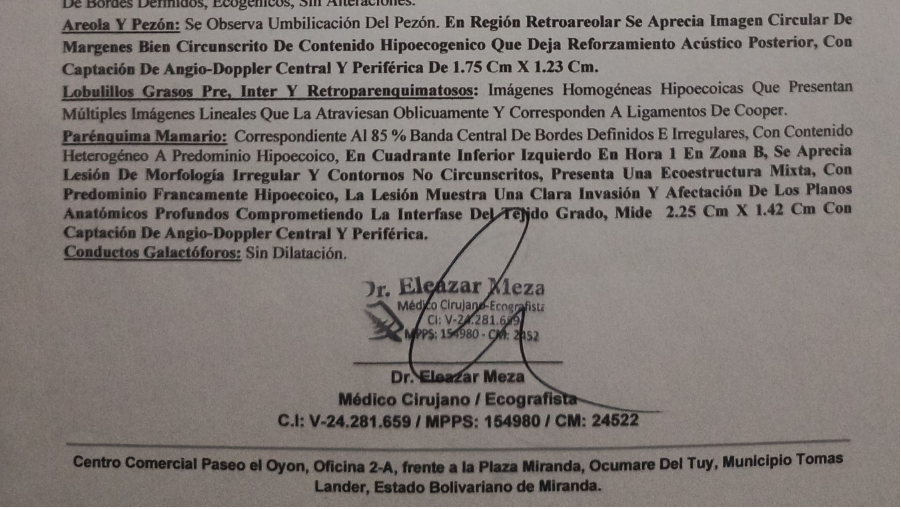
Dr. Eleazar Meza
Médico Cirujano / Ecografista
C.i: V-24.281.659 / MPPS: 154980 / CM: 24522
Fecha: 5/10/2026
Ecosonograma Mamario
Paciente: Margret Pena
Cedula: [CENSURADO]
Edad: 49 Años
Se Realiza Estudio Ecográfico Con Equipo Vinmoa3 Transductor Lineal 8-16 MHz
Mama Derecha: Ecotextura De Fondo Ecomixta Compuesta Por Tejido Graso Y Fibroglandular. Complejo Cutáneo De Bordes Defnidos, Ecogénicos, Sin Alteraciones
Areola Y Pezón: Se Observa Umbilicación Del Pezón, En Región Retroareolar Se Aprecia Imagen Circular De Margenes Bien Circunscrito De Contenido Anecogénico Que Deja Reforzamiento Acústico Posterior Sin Captación De Aurgi-Doppler Central Ni Periférica De 0.53 Cm X 0.49 Cm.
Lobulillos Grasos Pre, Inter Y Retroparenquimatosos: Imágenes Homogéneas Hipoeco Que Presentan Múltiples Imágenes Lineales Que La Atraviesan Oblicuamente Y Corresponden A Ligamentos De Cooper.
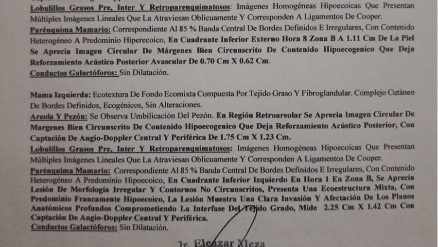
Parénquima Mamario: Corresoibduebte Al 85% Banda Central De Bordes Definidos E Irregulares, Con Contenido Heterogéneo A Predominio Hiperecoico, En Cuadrante Inferior Externo Hora 8 Zona B A 1.11 Cm De La Piel Se Aprecia Imagen Circular De Márgenes Bien Circunscrito De Contenido Hipoecogenico Que Deja
Reforzamiento Acústico Posterior Avascular De 0.70 Cm X 0.62 Cm.
Conductos Galactóforos: Sin Dilatación
Mama Izquierda: Ecotextura De Fondo Ecomixta Compuesta Por Tejido Graso Y Fibroglandular. Complejo Cutáneo De Bordes Definidos, Ecogénicos, Sin Alteraciones
Areloa
Areola Y Pezón: Se Observa Umbilicación Del Pezón, En Región Retroareolar Se Aprecia Imagen Circular De Margenes Bien Circunscrito De Contenido Hipoecogenico Que Deja Reforzamiento Acústico Posterior, Con Captación De Aurgi-Doppler Central Y Periférica De 1.75 Cm X 1.23 Cm.
Lobulillos Grasos Pre, Inter Y Retroparenquimatosos: Imágenes Homogéneas Hipoeco Que Presentan Múltiples Imágenes Lineales Que La Atraviesan Oblicuamente Y Corresponden A Ligamentos De Cooper.
Parénquima Mamario: Corresoibduebte Al 85% Banda Central De Bordes Definidos E Irregulares, Con Contenido Heterogéneo A Predominio Hiperecoico, En Cuadrante Inferior inferior Izquierdo En Hora 1 Zona B, Se Aprecia Lesión De morfologia Irregular Y Contornos No Circunseritos, Presenta Usa Ecoestructura Mixta, Con Anatómicos Profundos Comprometiendo La Interfase Del Tejido Grado, Mide 2.25 Cm x 1.42 Cm Con Captación De Angio-Doppler Central Y Periférica
Conductos Galactóforos: Sin Dilatación
English Transcript
Dr. Eleazar Meza
Physician / Sonographer
ID: V-24.281.659 / MPPS: 154980 / CM: 24522
Date: 5/10/2026
Breast Ultrasound
Patient: Margret Pena
ID: [REDACTED]
Age: 49 years
Ultrasound study performed using Vinmoa3 equipment with an 8-16 MHz linear transducer.
Right Breast: Mixed background echotexture composed of fatty and fibroglandular tissue. Skin complex with defined, echogenic borders; no abnormalities observed.
Areola and Nipple: Nipple retraction observed. In the retroareolar region, a circular image with well-circumscribed margins and anechoic content is visible; it exhibits posterior acoustic enhancement and shows no central or peripheral Doppler flow. Dimensions: 0.53 cm x 0.49 cm.
Pre-, inter-, and retroparenchymal fatty lobules: Homogeneous hypoechoic images traversed obliquely by multiple linear structures corresponding to Cooper's ligaments.
Breast Parenchyma: Corresponds to 85% [of the tissue]. Central band with defined, irregular borders and heterogeneous content (predominantly hyperechoic). In the lower outer quadrant (8 o'clock position, Zone B), at a depth of 1.11 cm from the skin, a circular image with well-circumscribed margins and hypoechoic content is observed; it exhibits posterior acoustic enhancement and is avascular. Dimensions: 0.70 cm x 0.62 cm. Galactophorous ducts: No dilation.
Left breast: Mixed background echotexture composed of fatty and fibroglandular tissue. Skin complex with defined, echogenic borders; no abnormalities.
Areola and nipple: Nipple retraction is observed. In the retroareolar region, a circular image with well-circumscribed margins and hypoechoic content is visible, exhibiting posterior acoustic enhancement and central and peripheral Angio-Doppler flow; it measures 1.75 cm x 1.23 cm.
Pre-, inter-, and retroparenchymal fatty lobules: Homogeneous hypoechoic images traversed obliquely by multiple linear structures corresponding to Cooper's ligaments.
Breast parenchyma: Central band with defined yet irregular borders and heterogeneous, predominantly hyperechoic content. In the lower-inner quadrant (left breast, 1 o'clock position, Zone B), an irregularly shaped lesion with non-circumscribed contours is observed; it presents a mixed echostructure and extends to deep anatomical structures, involving the fatty tissue interface. It measures 2.25 cm x 1.42 cm and shows central and peripheral Angio-Doppler flow.
Galactophorous ducts: No dilation.
Spanish Transcript:
Dr. Eleazar Meza
Médico Cirujano / Ecografista
C.i: V-24.281.659 / Mpps: 154980 / Cm: 24522
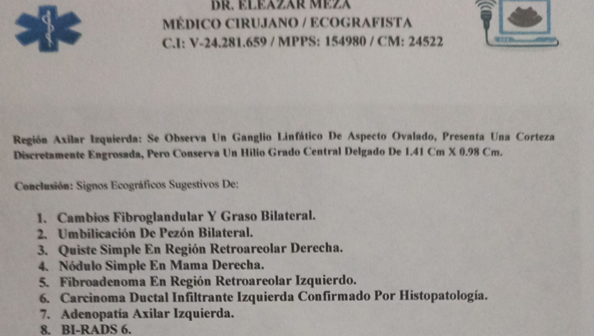
Región Axilar Izquierda: Se Observa Un Ganglio Linfático De Aspecto Ovalado, Presenta Una Corteza Discretamente Engrosada, Pero Conserva Un Hilio Grado Delgado De 1.41 Cm X 0.98 Cm.
Conclusión: Signos Ecográficos Sugestivos De:
1. Cambios Fibroglandular y Graso Bilateral.
2. Umilicación De Pezón Bilateral.
3. Quiste Simple En Región Retroareolar Derecha.
4. Nódulo Simple En Mama Derecha.
5. Fibroadenoma En Región Retroareolar Izquierdo.
6. Carcinoma Ductal Infiltrante Izquierda Confirmado Por Histopatología
7. Adenopatia Arilar Izquierda.
8. BI-RADS 6.
English Transcript:
Dr. Eleazar Meza
Physician and Surgeon / Sonographer
ID: V-24.281.659 / MPPS: 154980 / CM: 24522
Left Axillary Region: An oval-shaped lymph node is observed; it presents a mildly thickened cortex but retains a thin hilum, measuring 1.41 cm x 0.98 cm.
Conclusion: Ultrasound findings suggestive of:
1. Bilateral fibroglandular and fatty changes.
2. Bilateral nipple retraction.
3. Simple cyst in the right retroareolar region.
4. Simple nodule in the right breast.
5. Fibroadenoma in the left retroareolar region.
6. Left infiltrating ductal carcinoma (histopathologically confirmed).
7. Left axillary lymphadenopathy.
8. BI-RADS 6.
Spanish Transcript:
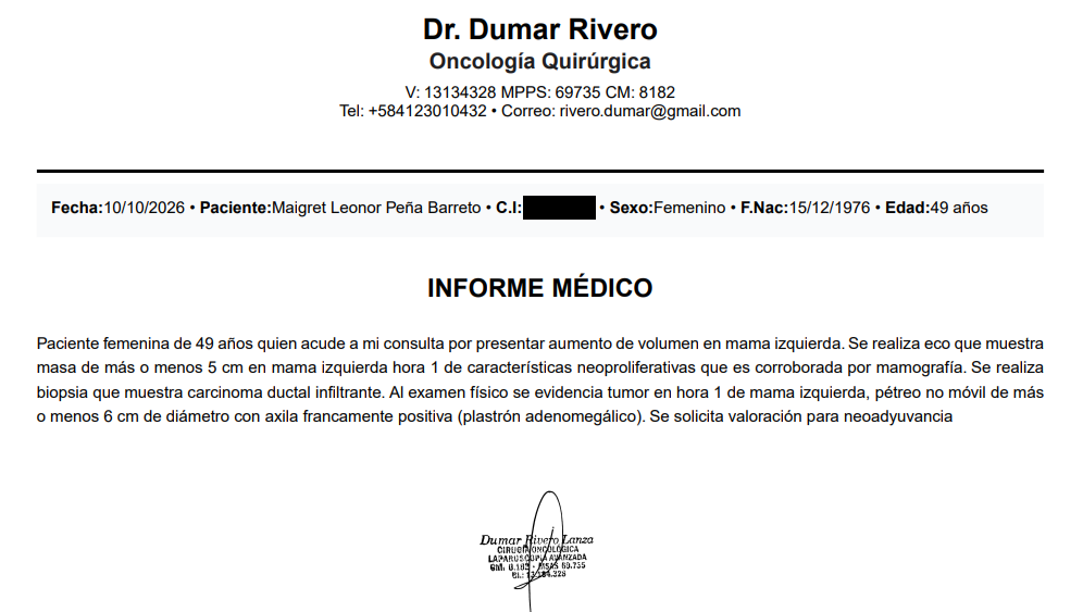
Dr. Dumar Rivero
Oncología Quirúrgica
V: 13134328 MPPS: 69735 CM: 8182
Tel: +584123010432 • Correo: [email redacted]
Fecha:10/10/2026 • Paciente:Maigret Leonor Peña Barreto • C.I:[CENSURADO] • Sexo:Femenino • F.Nac:15/12/1976 • Edad:49 años
INFORME MÉDICO
Paciente femenina de 49 años quien acude a mi consulta por presentar aumento de volumen en mama izquierda. Se realiza eco que muestra masa de más o menos 5 cm en mama izquierda hora 1 de características neoproliferativas que es corroborada por mamografía. Se realiza biopsia que muestra carcinoma ductal infiltrante. Al examen físico se evidencia tumor en hora 1 de mama izquierda, pétreo no móvil de máso menos 6 cm de diámetro con axila francamente positiva (plastrón adenomegálico). Se solicita valoración para neoadyuvancia
English Transcript:
Dr. Dumar Rivero
Surgical Oncology
V: 13134328 MPPS: 69735 CM: 8182
Tel: +584123010432 • Email: [email redacted]
Date: 10/10/2026 • Patient: Maigret Leonor Peña Barreto • ID No.: [REDACTED] • Sex: Female • DOB: 15/12/1976 • Age: 49 years
MEDICAL REPORT
A 49-year-old female patient presents with enlargement of the left breast. Ultrasound reveals a mass of approximately 5 cm in the left breast at the 1 o'clock position, displaying features suggestive of malignancy; this finding is confirmed by mammography. A biopsy is performed, revealing infiltrating ductal carcinoma. Physical examination reveals a tumor at the 1 o'clock position of the left breast—stony-hard and fixed, measuring approximately 6 cm in diameter—along with clear axillary involvement (conglomerate lymphadenopathy). Evaluation for neoadjuvant therapy is requested.
Organizer