- B
- S


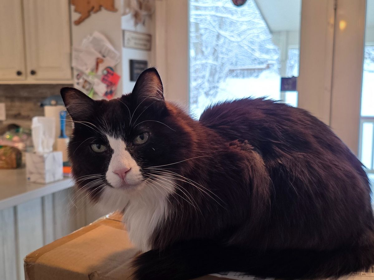
Hello, we are Emily and Nathan. We are the lucky owners of six amazing cats and about to give birth to our first human girl child in the next few days. Unfortunately, in November, our one female kitty, Gemma, started showing signs of very labored breathing and coughing. Eventually this turned into lethargy and not wanting to eat/drink.
We took her in to a local emergency vet here in the Valley and unfortunately it turned into a fiasco of events that almost ended her life. She was not getting the care that she needed (and in fact, actually got worse with their negligence and lack of treatment). We've since taken her to Pet ER in Anchorage where she has been for the last several days.
Since December she has been in and out of the vet numerous times, with hospitalizations dating:
11/29/2025-12/1/2025 in the Valley
1/7/2026-1/9/2026 in the Valley
3/9/2026-?/?/2026 in the Valley (her records from the location in the Valley are very confusing so this date range was hard for me to figure out)
4/2/2026-4/6/2026 in the Valley
4/6/2026-current in Anchorage
If you scroll to the bottom of this message, I've attached the initial summary of Gemma's history if you'd like to see what her current doctor has summarized.
We have been fortunate enough to have family that has been so incredibly gracious to help us out where we've needed it during this time but we have decided to try this out as a way to ask for additional help. As mentioned above, we're scheduled to have our first child this weekend and we have no idea what the future has in store for us as a new family or for Gemma but we want to do everything in our power to make sure that our baby girl kitty meets our baby human girl.
We love our cats as if they are our children, as any pet owner should, and we want to give Gemma the longest, happiest life that we can, just as we want for our new baby.
Any help is appreciated immensely - we thank you for reading this far!
Small Animal Internal Medicine CONSULTATION
Gemma is a 9-year 2-month-old spayed female domestic shorthair who presented to PET ER on 4/6/26 as a transfer for severe respiratory distress and progressive multifocal pulmonary disease of several months’ duration. Historical review indicates chronic respiratory signs dating back years, with intermittent episodes of labored breathing that were previously self-limiting and not treated. On 11/29/25, Gemma developed acute coughing, lethargy, inappetence, and increased respiratory effort. Thoracic radiographs at that time identified a caudodorsal and right middle peripheral alveolar pulmonary pattern with a questionable rounded structure in the right caudal lung lobe, mild pleural effusion versus pleuritis, and gastric distension consistent with aerophagia. The imaging appearance was considered unusual, raising concern for infiltrative disease such as abscess, granuloma, or neoplasia, with inflammatory and infectious disease also considered. Cardiogenic edema was considered less likely, and pulmonary hemorrhage was also listed as a differential. Advanced diagnostics including thoracic ultrasound, CT, bronchoscopy with airway cytology, and infectious disease screening were recommended at that time.
Follow-up radiographs on 12/23/25 demonstrated persistent but mildly improved bilateral caudodorsal pulmonary consolidation (right greater than left) with milder right middle lung lobe involvement. FIV/FeLV testing was negative. The patient was empirically treated with amoxicillin-clavulanate (Clavamox) without clinical improvement. A thoracic CT performed 1/8/26 revealed persistent multifocal pulmonary consolidation, most severe in the caudal lung fields, with concurrent nodular-like lesions suspected on prior imaging. There was no evidence of abscess formation. Mild to moderate tracheobronchial lymphadenopathy was present and considered reactive versus early neoplastic. The abdomen was unremarkable. Differentials at that time included pulmonary neoplasia (carcinoma, histiocytic sarcoma, other), atypical infectious bronchopneumonia (including Mycoplasma, fungal disease, or other), and endogenous lipid pneumonia. Specialist consultation and airway sampling (bronchoscopy with BAL or transtracheal wash) and/or ultrasound-guided pulmonary aspirates were recommended; however, referral to internal medicine was not pursued at that time.
Airway sampling was reportedly performed, though documentation suggests this was more consistent with an endotracheal wash rather than a bronchoscopy-guided BAL. There was no documentation regarding sterile technique, bronchoscopy visualization, or sampling site, raising concern that the sample was not representative and may have been contaminated. Cytology showed purulent to pyogranulomatous inflammation with occasional intracellular bacteria (bacilli and cocci), degenerate neutrophils, and macrophages, along with moderate numbers of smudged respiratory epithelial cells concerning for oropharyngeal contamination. Findings consistent with feline asthma or neoplasia were not observed. Culture yielded low growth of normal flora, further supporting contamination. No Mycoplasma PCR or culture was performed. An intrathoracic aspirate was also attempted but was hypocellular and nondiagnostic, containing primarily blood and rare mesothelial cells without evidence of inflammation, infection, or neoplasia.
The patient was subsequently treated with doxycycline for 14 days and prednisolone (dose and taper not documented), with reported clinical improvement. Prednisolone was tapered and fluticasone initiated; however, respiratory signs recurred following steroid reduction.
Gemma represented on 4/2/26 with respiratory decompensation. CBC at that time showed hypokalemia (3.2), marked neutropenia (0.09), monocytosis (1.0), low eosinophils (0.16), and thrombocytopenia (95). Thoracic radiographs showed mild progression of multifocal pulmonary consolidation. Differentials prioritized atypical infectious pneumonia, with neoplasia and lipid pneumonia also considered. A lung aspirate performed at that time showed neutrophilic inflammation with epithelial cell proliferation; however, inflammation limited interpretation for neoplasia, and additional testing including Mycoplasma PCR was recommended. The patient was discharged but represented on 4/4/26 for worsening respiratory distress.
Patient was presented to PET ER in anchorage for IM consultation - Repeat imaging identified relatively static caudal and left cranial alveolar disease but progressive right cranial and right middle lung lobe consolidation, along with development of a pneumothorax suspected to be iatrogenic secondary to recent pulmonary FNA. Additional findings included gastric gas distension compatible with aerophagia. Chemistry revealed hyperglobulinemia. Due to the pneumothorax, advanced diagnostics such as repeat CT, bronchoscopy, BAL, respiratory PCR testing, and additional aspirates were deferred. Therapeutic thoracocentesis was recommended with monitoring for resolution, with consideration of chest tube placement if persistent. Pending stabilization, recommended diagnostics included toxoplasma titers, cryptococcal antigen testing, and consideration of abdominal imaging with FNA. If pneumothorax persists or disease progresses, thoracotomy with lung biopsy for histopathology and aerobic, fungal, and Mycoplasma cultures is recommended. If pneumothorax resolves, advanced diagnostics including CT, bronchoscopy, BAL cytology, aerobic/anaerobic/fungal cultures, and Mycoplasma PCR are recommended through the internal medicine service.
Overall, this case represents a chronic progressive multifocal pulmonary disease with intermittent steroid responsiveness, poor response to empirical antibiotics, nondiagnostic airway sampling, and progressive consolidation with nodular components. The leading differential diagnoses include atypical infectious pneumonia (particularly Mycoplasma, toxoplasmosis, cryptococcosis, fungal disease, or less likely lungworm), endogenous lipid pneumonia (possibly secondary to chronic pneumocyte injury and airway obstruction), and pulmonary neoplasia (including lymphoma, histiocytic sarcoma, diffuse carcinoma, or metastatic disease). Secondary inflammatory fibrosis may also be contributing. The prior steroid responsiveness does not exclude infection or neoplasia and may transiently improve inflammation associated with any of these etiologies. The previously reported purulent inflammation with intracellular bacteria may represent true infection but is confounded by likely contamination and lack of directed culture/PCR testing.
This patient should ideally have been referred to an internal medicine specialist in December 2025 when persistent caudodorsal consolidation and suspected pulmonary nodule were first identified, as earlier bronchoscopy-guided BAL, CT-guided sampling, and infectious disease testing may have provided a definitive diagnosis prior to disease progression and repeated invasive sampling.
Current recommended diagnostic plan includes continued monitoring and management of the iatrogenic pneumothorax with therapeutic thoracocentesis as needed and consideration of chest tube placement if persistent. While awaiting stabilization, serum toxoplasma titers and cryptococcal antigen testing are recommended. Once pneumothorax resolves, repeat thoracic CT followed by bronchoscopy with bronchoalveolar lavage is recommended, with samples submitted for cytology, aerobic/anaerobic culture, fungal culture, Mycoplasma PCR, respiratory PCR panel, and evaluation for lipid-laden macrophages. If bronchoscopy is nondiagnostic or disease continues to progress, surgical lung biopsy via thoracotomy is recommended for definitive histopathology and culture. Abdominal imaging with FNA of any abnormal organs may also be considered given hyperglobulinemia and concern for systemic disease.
While awaiting advanced diagnostics, medical management should include continuation of pradofloxacin and clindamycin to provide coverage for atypical bacterial and protozoal infections, discontinuation of doxycycline to avoid redundant antimicrobial therapy, and continuation of prednisolone at approximately 0.5 mg/kg PO once daily to control inflammatory pulmonary injury, particularly if endogenous lipid pneumonia or inflammatory pneumonitis is contributing. Oxygen support and careful monitoring for worsening pneumothorax are recommended. Recommend completion of a 50 mg/kg PO SID coarse of fenbendazole.
Prognosis remains guarded to poor and depends heavily on the underlying etiology. If an atypical infectious etiology is identified and responsive to therapy, prognosis may be fair with prolonged treatment. Endogenous lipid pneumonia carries a guarded prognosis, particularly when diffuse and chronic, though some cats stabilize with anti-inflammatory therapy. Pulmonary neoplasia carries a poor prognosis, particularly if diffuse or multifocal, with survival often measured in weeks to a few months. The presence of progressive multifocal consolidation, recurrence after steroid taper, hyperglobulinemia, cytologic inflammation, and worsening clinical signs despite empirical therapy collectively suggest advanced pulmonary disease, and risk of further respiratory decompensation remains high.
PENDING
Toxoplasma IFA pending
Cryptococcus agglutination antigen pending
Cytology - pending
TO DO
Abdominal Ultrasound
+/- FNA
Disclaimer:
Conclusions are based on the history and information available to the clinician at the time of consultation. Patients receiving internal medicine consultations are not considered established clients of the Internal Medicine Service. If ongoing care with the Internal Medicine Service is desired, please submit a referral at: https://www.petemergencyak.com/referring-internal-medicine --
Dr. Ashley Melco, DVM, DACVIM (SAIM)
Small Animal Internal Medicine Specialist
PET ER - Specialty Service
President of the Alaska Veterinary Medical Association
Pet Emergency Treatment
Internal Medicine Service
2320 E Dowling Rd
Anchorage AK 99507
907-274-5636