- W
- W
- J
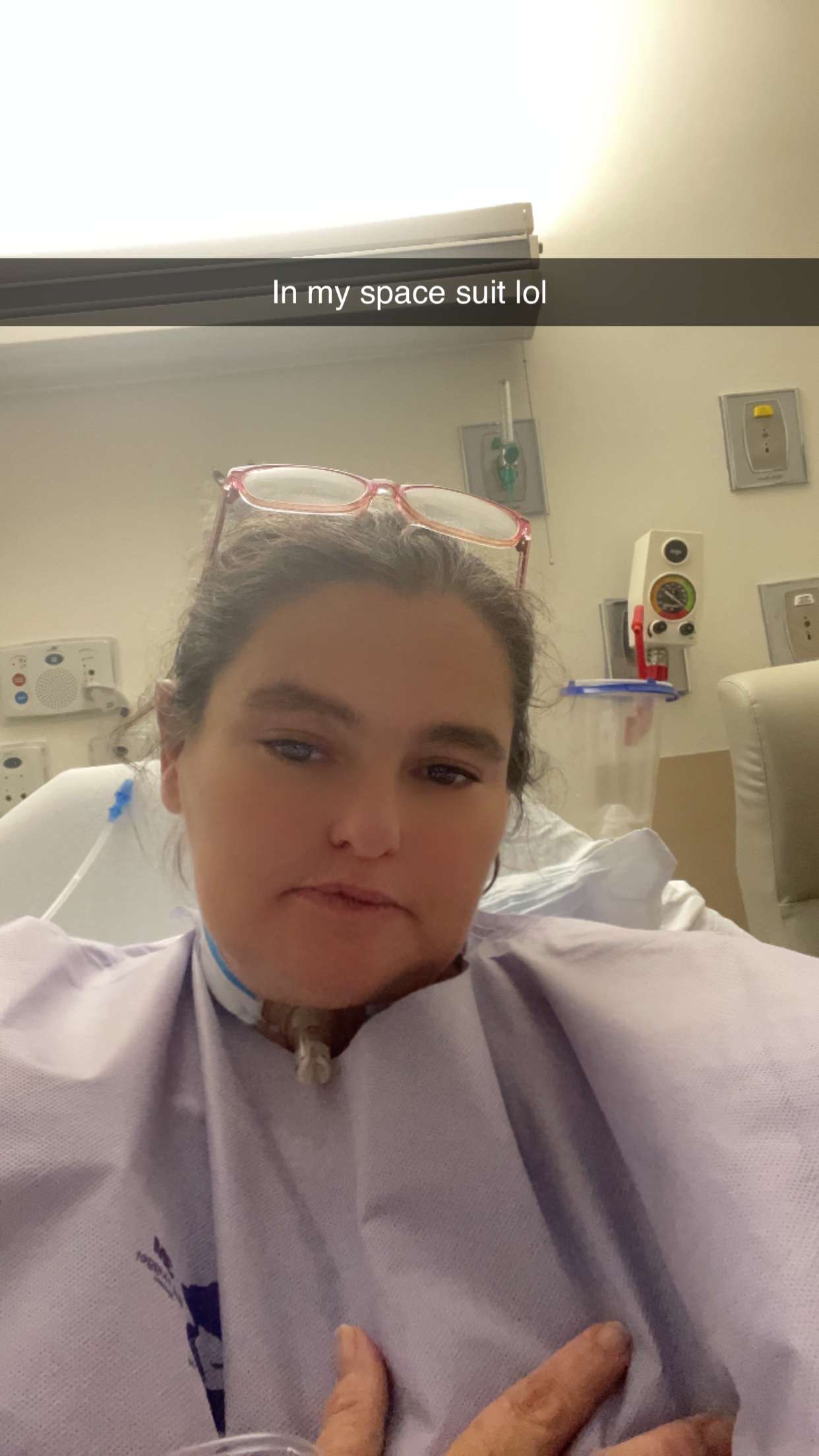
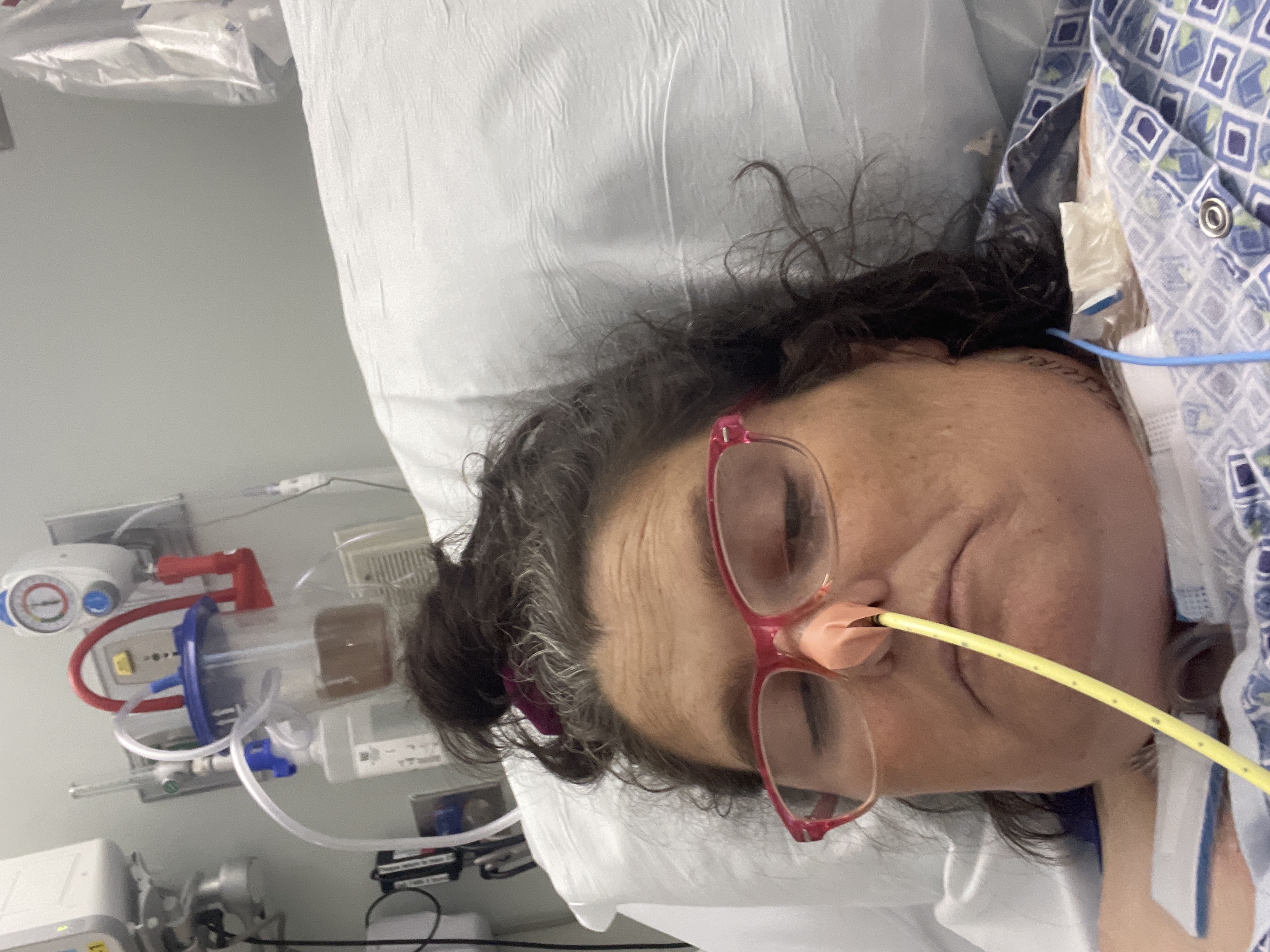
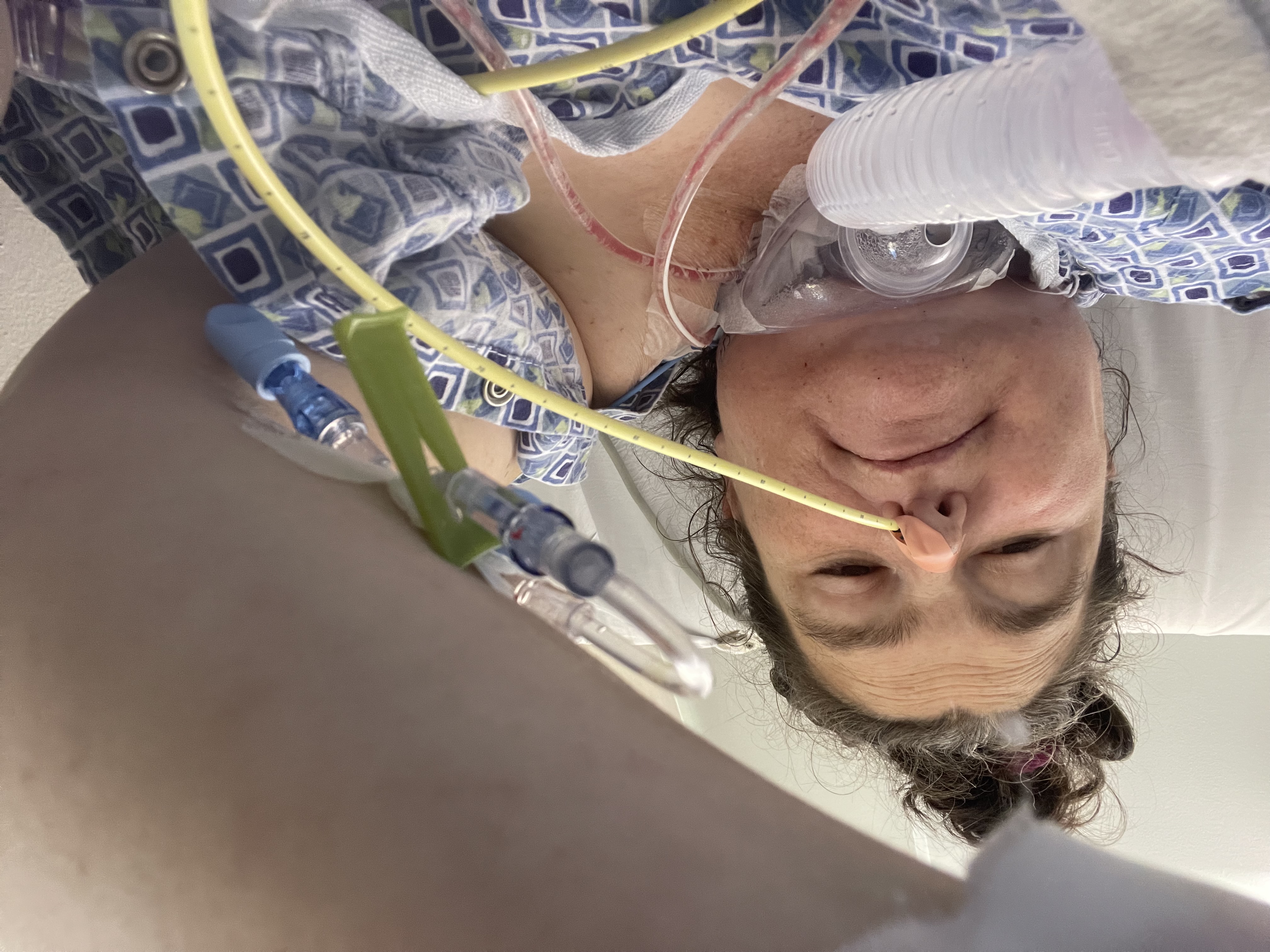
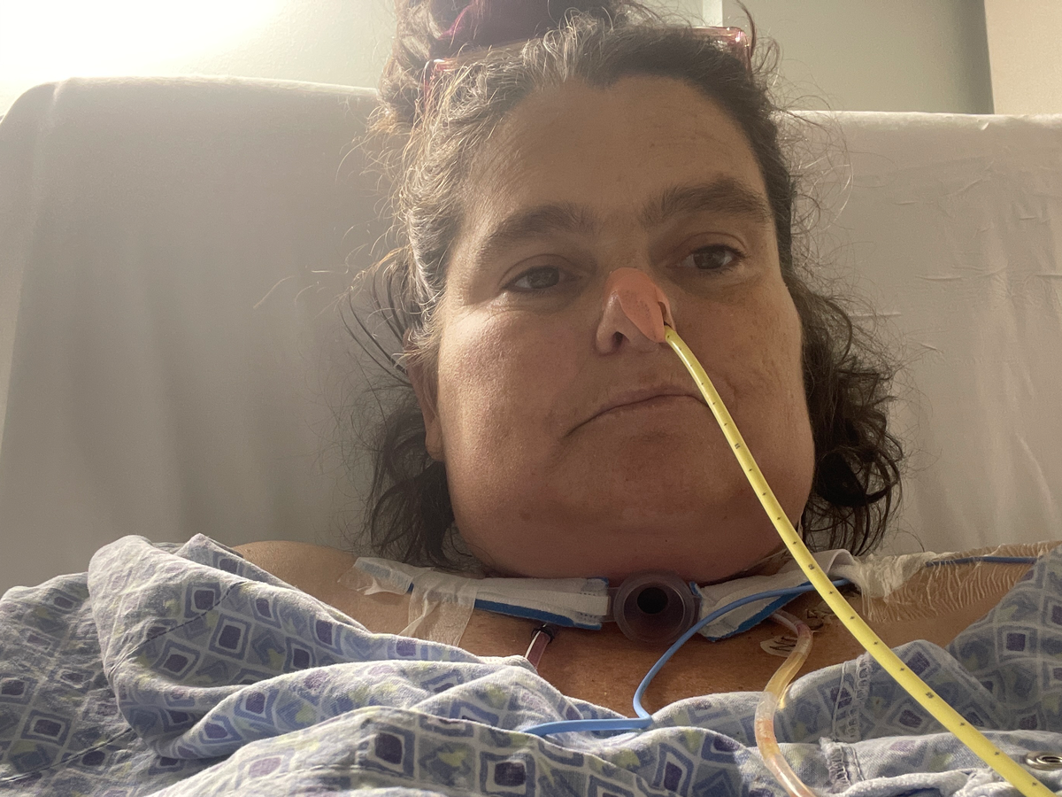
My name is Christy. I am 44 years old. About 6-9 months ago, I had major issues getting my voice to be normal. So, I went to the doctor, and they had me go see an ENT doctor. At this point, he found what he believed was a tumor on my voice box. But to be sure, he took a biopsy of my right lymph node where he saw it had spread so far. The biopsy was sent out and came back positive for larynx cancer of the throat. We put in for more tests to decide on the best treatment, like just needing radiation or having to go in and remove it along with my voice box/vocal cords. After tests, we came to the agreement that I should have the surgery. The cancer had spread to my right lymph node and was blocking my airway as well as my swallow gland area. So, it was important to go in and get it all out. I wasn't very okay with this option, but to stay alive, it's what I had to do. My surgery couldn't happen for a month and a half, so we had to first do a tracheotomy to help me breathe until the surgery. That went okay as it couldn't happen. Then March 17th was here, time for my surgery. I came in at 5 am and started surgery around 7 am. It lasted 11 hours. I woke up super confused. This is the note from my doctor:
PROCEDURES PERFORMED: Total laryngectomy, bilateral neck dissection, free flap reconstruction of the hypopharynx. This is a combined case with Dr. Mokhtari. My portion of the procedure was the total laryngectomy and bilateral neck dissection. Dr. Mokhtari will separately dictate the reconstruction and with free flap reconstruction of the hypopharynx. The patient was brought to the operating room and under anesthesia through the previously placed tracheotomy site, the patient's neck was prepped with Betadine and draped for total laryngectomy and neck dissections. In addition, the right arm was prepped for a donor site for a radial forearm free flap reconstruction. A curved incision was made from the right mastoid curving down along the left neck and then across at the previous trach site and then onto the left neck. Skin flap was elevated superiorly to the level of the hyoid bone bilaterally. The incision was then carried around the previous trach site to isolate this and permit the lower skin flap to be elevated down to the clavicle and sternum. First, a right neck dissection was performed. Dissection was carried down to the sternocleidomastoid muscle. The retromandibular vein and greater auricular nerve were divided and the sternocleidomastoid muscle was retracted posteriorly. There was evidence of invasion of the metastatic nodes into the medial aspect of the sternocleidomastoid muscle. Therefore, a portion of the medial aspect of the sternocleidomastoid muscle was left attached to the nodes. The remainder of the sternocleidomastoid muscle was retracted laterally identifying the spinal accessory nerve, which was preserved, as the muscle was retracted. Dissection was then carried medial to the sternocleidomastoid muscle down to the cervical plexus. The cervical plexus branches were followed anteriorly into the neck, elevating the nodes in the jugular chain down to the supraclavicular nodes, which were then elevated and carried forward toward the jugular vein. The jugular vein was identified inferiorly and the node-bearing tissue was then elevated off the jugular vein from inferior proceeding superiorly. At approximately the midportion of the neck, it was apparent that the nodes in the right neck were adherent to the jugular vein. Dissection of the nodes off the jugular vein would not be satisfactory. Therefore, the jugular vein was ligated inferiorly and superiorly and taken along with the adenopathy. The dissection was then carried medially to the carotid artery. The nodes were elevated off the carotid artery and vagus nerve. Dissection was then carried superiorly to identify the hypoglossal nerve, which was then followed into the tongue and preserved, carrying the dissection superiorly to the level of the digastric muscle and then forward to the level of the submandibular gland. The facial vein was ligated as it passed into the submandibular gland. The submandibular gland was retracted superiorly and this permitted exposure of the larynx. The dissection was carried down to the hyoid bone. The hyoid bone was freed from the tongue base, carrying the dissection along the greater corner of the hyoid bone bilaterally. It was apparent that there was a tumor expanding from the larynx into the paralaryngeal space on the right side. The nodes of the neck were left attached to the larynx at this point. Inferiorly, the strap muscles were divided, carrying the dissection down to the trachea. The thyroid isthmus was divided leaving the right thyroid lobe attached to the specimen and elevating the left thyroid lobe off the trachea. On the left side, the dissection was carried through the omohyoid muscle. The pharyngeal constrictor muscles were divided carrying the dissection down to the parathyroid cartilage. It was possible to enter the larynx on the left into the piriform sinus and then across the tongue base and then rotating the larynx to the right. The pharyngeal mucosa was incised along the lateral pharyngeal wall on the left and then carrying the dissection inferiorly to the esophagus. The resected mucosa was primarily on the left side, preserving the mucosal strip between the hypopharynx and the esophagus. This permitted the specimen to be removed and this included the left piriform sinus, total laryngectomy, and lymph nodes in levels 2, 3, and 4. Bleeding was controlled with electrocautery and silk ties. Margins were taken around the pharyngeal resection and these were all negative for tumor. Because of the resection of the jugular vein, it was necessary to perform a left neck dissection primarily for clearance of the lymph nodes and obtaining recipient vessels for the planned free flap reconstruction. On the left side, dissection was carried medial to the sternocleidomastoid muscle. Node-bearing tissue was elevated off the jugular vein and carotid artery and dissection was then carried superiorly including nodes in levels 2, 3, and 4, and finally completing the dissection by rotating the node-bearing tissue off the jugular vein and exposing the facial vein as a recipient vessel for the reconstruction. The nodes at levels 2, 3, and 4 were removed and sent for histologic diagnosis. Bleeding was controlled with electrocautery and silk ties. At this point, Dr. Mokhtari proceeded with the reconstruction of the hypopharynx with a free flap and her portion of the procedure will be dictated separately.
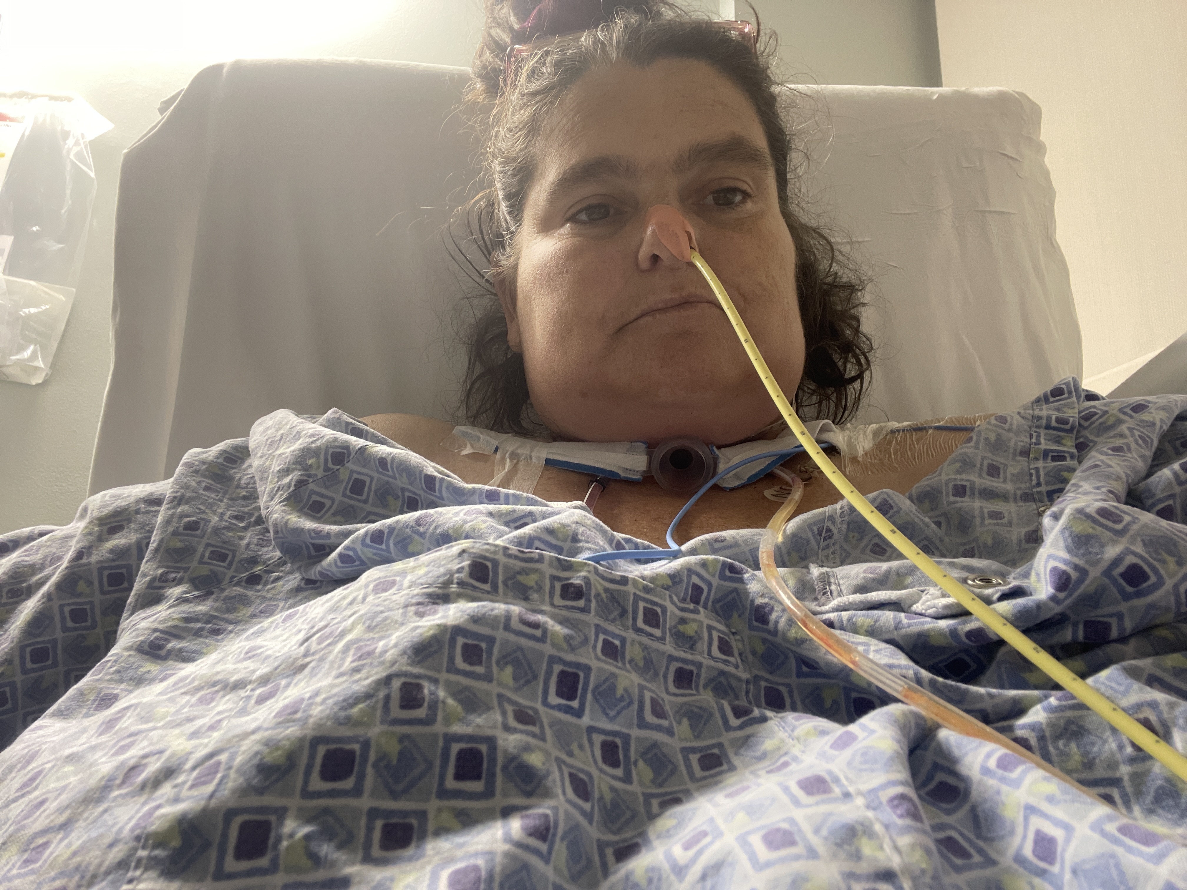
So yeah, that's everything. I'm now 1 week and 1 day since surgery. I am going to have to do some radiation and chemotherapy to make sure nothing was left behind .I'm hoping to go home soon. I didn't want to make a GoFundMe, but the money for travel and things needed that my insurance won't cover is just going to be too much. If you can help, please do. If not, I understand and am just grateful for u all and that I am still alive. Love you all so much.